Ohio State University's Wexner Medical Center is the first in the U.S. to install new Magnetic Resonance Imaging (MRI) developed by Siemens in collaboration with the OSU medical school researchers, a news release said.
The new device is designed for patients who have implanted medical devices, severe obesity or claustrophobia.
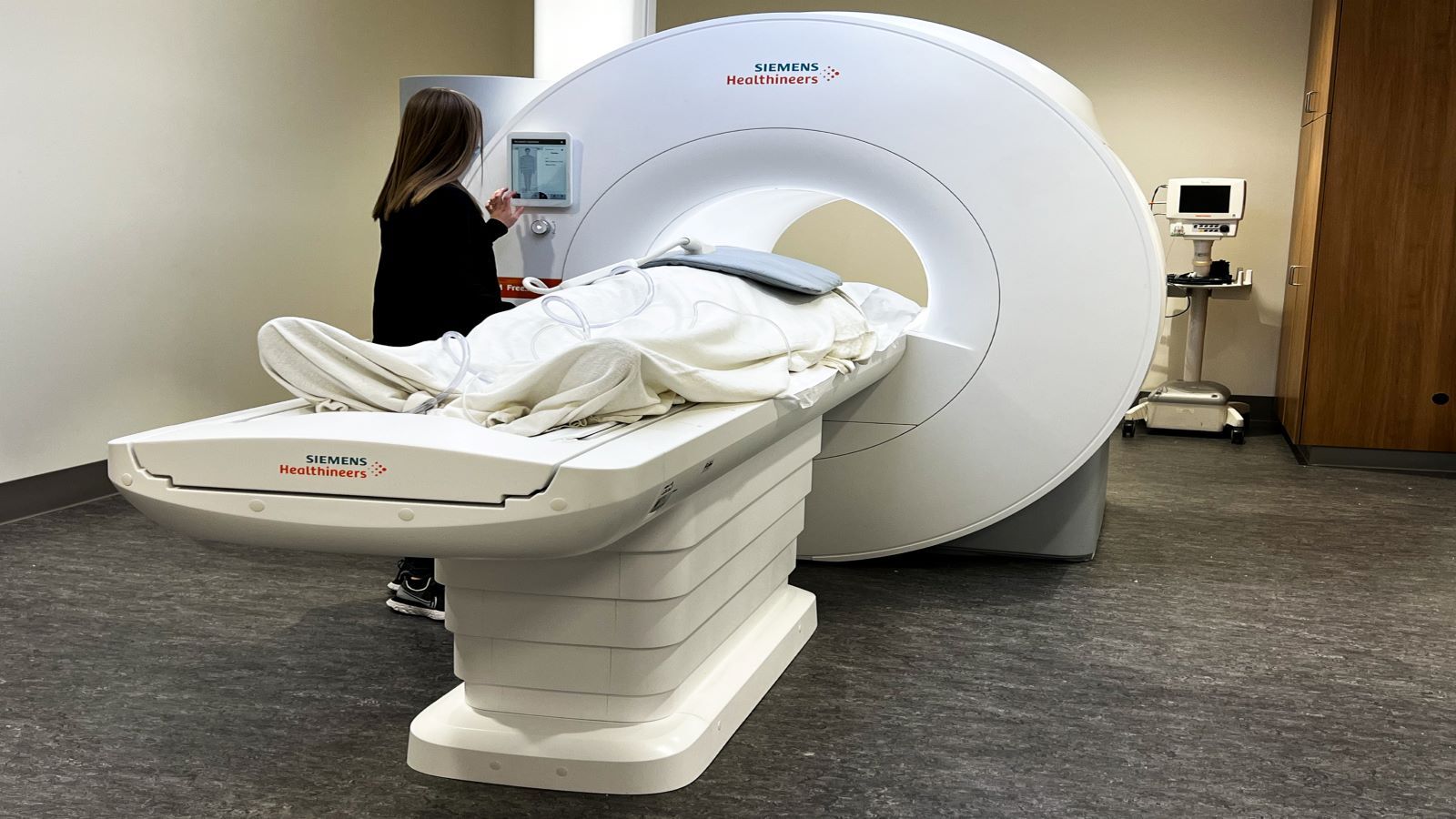
"The new machine has the largest MRI opening to date, 80 centimeters, compared to the typical 60-70 centimeters, and a lower magnetic field strength that offers the potential for it to be used for lung imaging without X-ray radiation," the news release said.
MRIs are primary used to take images of the brain, spine and joints but can also used for heart and blood vessel imaging.
“Many of our patients have pacemakers or defibrillators, and while many of those devices are now safe for MRI scanning, the metal in them can distort the magnetic field and corrupt the image quality," Orlando Simonetti, OSU research director of cardiovascular magnetic resonance said in the release. "We were looking for ways to improve the quality of images in these patients, and lower magnetic field strength could offer an advantage."
Simonetti worked with Rizwan Ahmad, an assistant professor of biomedical engineering at Ohio State, to suppress interference in the images, and produce clearer images at lower field strength, sharing their research with Siemens and contributing to the 0.55T Free.Max scanner.
“There’s no doubt in my mind that low-field MRI will play an important role in the future and will become more mainstream,” Simonetti said. “Going to lower field can reduce the cost of MRI systems and installation considerably, and with modern techniques for scanning and image processing, we can overcome the inherent loss of signal.”
OSU is also working with Nationwide Children’s Hospital to study how the new 0.55T scanner could be used with heart catherization.
"Children with congenital heart disease must undergo repeated heart catheterizations throughout their lives, and they are exposed to radiation every time they have an X-ray to guide the tube through a blood vessel to the heart," the news release said.